What is a Electromyography (EMG)?
Electromyography (EMG) is a diagnostic procedure to assess the health of muscles and the nerve cells that control them (motor neurons). EMG results can reveal nerve dysfunction, muscle dysfunction or problems with nerve-to-muscle signal transmission.
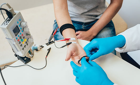
Motor neurons transmit electrical signals that cause muscles to contract. An EMG uses tiny devices called electrodes to translate these signals into graphs, sounds or numerical values that are then interpreted by a specialist.
During a needle EMG, a needle electrode inserted directly into a muscle records the electrical activity in that muscle.
How Do I Prepare for an EMG?
- Bathing: Take a shower or bath shortly before your exam in order to remove oils from your skin. Don’t apply lotions or creams before the exam.
- We will need to know if you have certain medical conditions. Tell the neurologist and other EMG lab personnel if you:
- Have a pacemaker or any other electrical medical device
- Take blood-thinning medications
- Have hemophilia, a blood-clotting disorder that causes prolonged bleeding
What Should I Expect During an EMG?
Before the procedure
You’ll likely be asked to change into a hospital gown for the procedure and lie down on an examination table. To prepare for the study, the neurologist or a technician places surface electrodes at various locations on your skin depending on where you’re experiencing symptoms. Or the neurologist may insert needle electrodes at different sites depending on your symptoms.
During the procedure
When the study is underway, the surface electrodes will at times transmit a tiny electrical current that you may feel as a twinge or spasm. The needle electrode may cause discomfort or pain that usually ends shortly after the needle is removed.
During the needle EMG, the neurologist will assess whether there is any spontaneous electrical activity when the muscle is at rest — activity that isn’t present in healthy
muscle tissue — and the degree of activity when you slightly contract the muscle.
He or she will give you instructions on resting and contracting a muscle at appropriate times. Depending on what muscles and nerves the neurologist is examining, he or she
may ask you to change positions during the exam.
If you’re concerned about discomfort or pain at any time during the exam, you may want to talk to the neurologist about taking a short break.
What Happens After the EMG?
After the MRI is complete you are free to leave and resume your normal activities.