
Imaging with room to breathe.
MRI, CT and X-ray at five Houston locations. Our MRI service includes open systems — magnet above and below, sides left open — for patients who need the extra room.
Five locations across Houston and Sugar Land. Bring an order from your doctor, or have them fax it to (888) 663-9778.
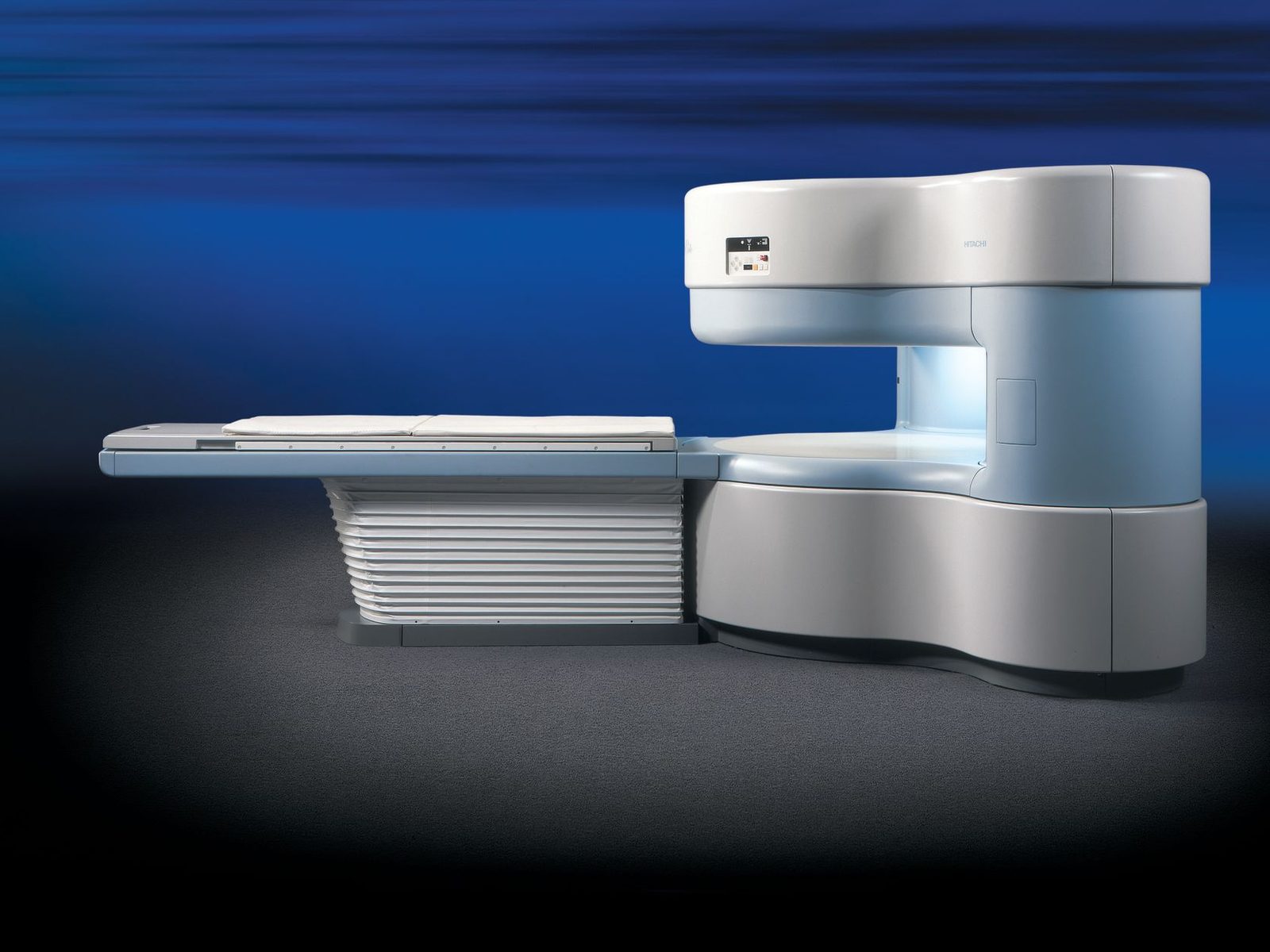
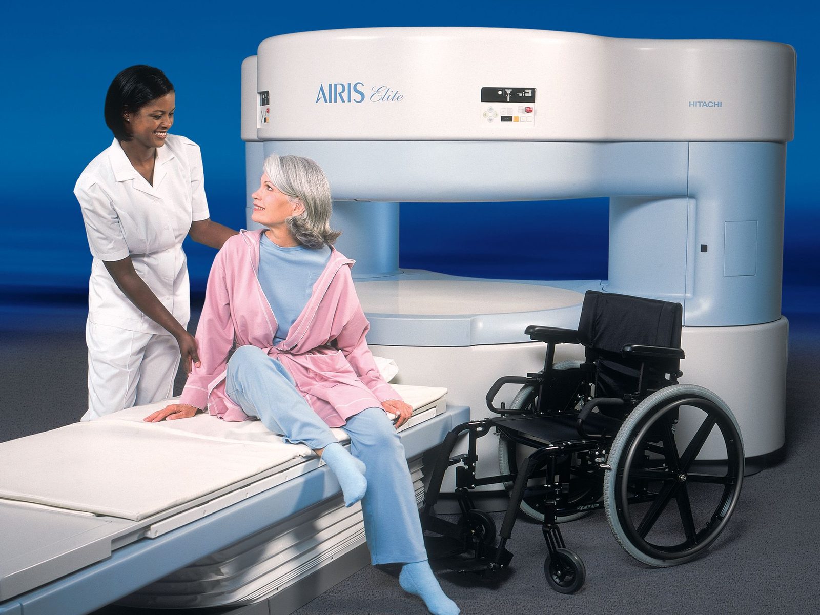
Open MRI. One of the MRI options we offer. The magnet sits above and below only, so you keep a clear line of sight to the room and there’s space beside the table for a family member.
Three ways to see what's going on.
Your doctor's order says which one you need. If you're not sure what you're scheduled for, bring the order or have the office fax it and we'll sort it out.
MRI
Detailed images of soft tissue — discs and spinal cord, knees, shoulders, brain, ligaments and tendons. Uses a magnetic field and radio waves, not radiation.
MRI → Fast and detailedCT scan
Cross-sectional images built from X-rays. Strong for bone detail, chest and abdomen, sinuses, kidney stones, and anything that needs answering quickly.
CT scan → In and outX-ray
The quickest look at bones and chest — fractures, joints, alignment, post-injury follow-up. Nothing to prepare, and you stay dressed unless we ask otherwise.
X-ray →What an MRI shows, part by part.
An MRI reads how tissue responds to a magnetic field, so it picks up things no other exam resolves. Select an area to see what shows up there.
Select a region
Tap a point on the diagram, or pick from the list, to see what an MRI picks up in that part of the body and how long the scan usually runs.
More about the examMRI separates grey matter from white matter and shows soft tissue the skull hides on other exams.
What the images show- Stroke and older infarcts
- Tumors and lesions
- Demyelination
- Pituitary and inner ear
- Vessels, on an MRA study
The one exam that shows the discs, the nerve roots and the spinal cord itself in the same series.
What the images show- Disc herniation and bulging
- Nerve root compression
- Cord compression
- Canal and foraminal narrowing
- Ligament injury after whiplash
Soft tissue around the joint — the structures that fail long before the bone does.
What the images show- Rotator cuff tears
- Labral tears
- Biceps tendon
- Bursitis and impingement
- Cartilage wear
MRI is used selectively here, for questions about soft tissue rather than the lungs themselves.
What the images show- Chest wall and mediastinum
- Brachial plexus
- The heart, on a cardiac study
- Soft-tissue masses
The stretch of spine behind the ribcage, where the cord is at its most confined.
What the images show- Disc herniation
- Cord compression
- Compression fractures
- Marrow changes from infection or tumor
- Canal narrowing
Tendon and ligament detail that plain films can't resolve.
What the images show- Ligament tears
- Tennis and golfer's elbow
- Loose bodies
- Cartilage damage
- Ulnar nerve
High contrast between organs and the tissue around them, without radiation.
What the images show- Liver lesions
- Bile ducts, on an MRCP study
- Pancreas
- Kidneys and adrenal glands
- Bowel inflammation
The most requested MRI we perform — and the exam that usually explains sciatica.
What the images show- Disc herniation and bulging
- Nerve root impingement
- Canal and foraminal stenosis
- Facet joint arthritis
- Slipped vertebra
- Marrow and endplate changes
Small structures at high resolution, including fractures that don't appear on an X-ray.
What the images show- Cartilage and TFCC tears
- Occult scaphoid fracture
- Ligament and tendon tears
- Ganglion cysts
- Loss of blood supply to bone
Bone marrow and deep soft tissue, where hip pain is often coming from something the X-ray missed.
What the images show- Labral tears
- Loss of blood supply to the femoral head
- Stress and hidden fractures
- Muscle and tendon tears
- Bursitis
- Sacroiliac joints
Every soft-tissue structure in the joint, in one series.
What the images show- Meniscal tears
- ACL and PCL tears
- Collateral ligaments
- Cartilage loss
- Bone bruising
- Baker's cyst
Ligament, tendon and bone detail after a twist, a fall, or pain that hasn't settled.
What the images show- Ligament tears
- Achilles and peroneal tendons
- Plantar fascia
- Stress fractures
- Cartilage and bone lesions
- Nerve thickening
Your images are interpreted by a radiologist alongside your history and symptoms. This describes what an MRI is capable of showing in each area — it isn't a diagnosis, and it doesn't replace your doctor's assessment.
Five steps, and we handle three of them.
- 01
The order comes in
Your doctor faxes it to (888) 663-9778 or emails info@originmri.com. You can also bring a paper order in.
- 02
We call you
We confirm what the exam needs, answer your questions, and pick a location and time that works.
- 03
Safety screening
A short form about implants, metal, prior surgeries and pregnancy. This one matters — please answer it carefully.
- 04
The scan
You'll hear knocking sounds while images are taken. We talk to you between sequences and you hold a call button.
- 05
Results to your doctor
A radiologist reads the images and sends a report to whoever ordered the scan.
Mild sedation is available on site. All medication is provided solely at the clinical discretion of our medical provider following an in-person health screening. Ask when you book, and arrange a ride home in case it is given.
Pick the freeway you're already on.
Same phone number for all five. Tap a pin to see the address, or call and we'll book you wherever is easiest to reach.
Schematic — not to scale
Choose a location
Tap a pin or a name to see the address and directions. All five share one scheduling line, so you can also just call and we'll place you.
Katy Freeway
9135 Katy Freeway, Suite 102Houston, TX 77024
Memorial, Spring Branch, Bunker Hill, Hedwig Village, the Energy Corridor and the near west side.
Get directions →Tomball Parkway
19610 Tomball Parkway, Suite 300Houston, TX 77070
Willowbrook, Champions, Cypress, Tomball, Spring and the FM 1960 corridor.
Get directions →Gulf Freeway
10761 Gulf Freeway, Suite AHouston, TX 77034
South Houston, Pasadena, Hobby Airport, Pearland and the southeast side.
Get directions →North Freeway
7000 North Freeway, Suite 900AHouston, TX 77076
Northline, Aldine, Independence Heights, Greenspoint and the near north side.
Get directions →Sugar Land
17227 W Grand Parkway SSugar Land, TX 77479
Sugar Land, Missouri City, Richmond, Rosenberg, Riverstone and Fort Bend County.
Get directions →Send the order. We'll take it from there.
Fax or email the order and we'll contact your patient directly to complete MRI safety screening and schedule at whichever of our five sites is closest to them.
Patients who have failed a closed-bore study are welcome — that's the case our open MRI exists for.
- Scheduling
- (832) 982-0845
- Fax orders to
- (888) 663-9778
- info@originmri.com
- Modalities
- MRI · CT · X-Ray
- Sites
- 5 across Houston & Sugar Land
Accredited in MRI and CT.
Origin is an accredited facility of the Intersocietal Accreditation Commission for both MRI and CT. Accreditation means our equipment, staff qualifications, image quality and safety procedures have been reviewed against published national standards by an independent body — not simply claimed.
The IAC is one of the organizations designated by the Centers for Medicare & Medicaid Services to accredit suppliers of advanced diagnostic imaging.
Before you come in.
I panicked in a closed MRI before. Will this be different?
Physically, yes — the sides are open, so nothing surrounds your head and you can see out of the machine in both directions. Tell us about the previous attempt when you call so we can plan for it: extra time, someone at the table with you, and breaks between sequences.
Will I fit?
Open systems don't have a fixed tunnel to squeeze into, which is why they suit larger patients. There are still table limits that vary by scanner, so call with your height and weight and we'll confirm the right location before you make the trip.
Can my husband, wife or parent stay in the room?
Usually, yes — as long as they pass the same metal screening you do and there's no contraindication. Let us know when you book so we can screen them ahead of time instead of at the door.
How long will I be there?
An MRI is generally 20 to 45 minutes depending on the body part and whether contrast is ordered. A CT is usually under 15 minutes and an X-ray is quicker still. Plan on arriving about 15 minutes early for paperwork.
How loud is it?
An MRI makes knocking and buzzing sounds while images are being taken — that's the machine working, not a problem. We provide ear protection, and we talk to you between sequences so you always know how much is left.
Ready to get scheduled?
Call and we'll confirm what your exam needs and book you at whichever location is easiest to reach.